Shoulder Ligament Anatomy Diagram / A Survey Of Human Shoulder Functional Kinematic Representations Springerlink : This page is about shoulder anatomy ligaments and muscles,contains soft tissues of the shoulder,shoulder joint;
Shoulder Ligament Anatomy Diagram / A Survey Of Human Shoulder Functional Kinematic Representations Springerlink : This page is about shoulder anatomy ligaments and muscles,contains soft tissues of the shoulder,shoulder joint;. The shoulder joint (glenohumeral joint) is a ball and socket joint between the scapula and the humerus. You can see it enclosing the glenohumeral joint and you can see its attachment on the anatomical you've got the transverse humeral ligament and the coracohumeral ligament. Ligaments appear as crisscross bands that attach bone to bone and help stabilize joints. The transverse humeral ligament is not shown on this diagram. A joint capsule is a watertight sac that surrounds a joint.
The shoulder anatomy includes the anterior deltoid, lateral deltoid, posterior deltoid, as well as the 4 rotator cuff muscles. All about the shoulder muscles. The primary function of the shoulder girdle is to give strength and range of motion to the arm. The muscular system anatomical chart muscle anatomy human, shoulder joint anatomy chart, details about knee joint anatomy understanding arthritis anatomical chart. Static:gh ligaments, labrum & capsule and dynamic constraints:
Shoulder Anatomy Springerlink from media.springernature.com Last update february 25, 2021. Although the joint is held together by these extensive ligament and muscle attachments, certain types of forces can weaken the shoulder easily. The ligament connecting the coracoid process and the acromion is called the coracoclavicular ligament. Get a 20.000 second shoulder ligaments anatomy stock footage at 30fps. Notice superior labrum and attachment of the superior glenohumeral ligament. An image depicting shoulder anatomy can be seen below. There are many shoulder ligaments which each play an important role in shoulder joint stabilization to various degrees: The shoulder anatomy includes the anterior deltoid, lateral deltoid, posterior deltoid, as well as the 4 rotator cuff muscles.
Shoulder joint of human body anatomy infographic diagram with all parts including bones ligaments muscles bursa cavity capsule cartilage membrane for medical science education and health care.
The shoulder anatomy includes the anterior deltoid, lateral deltoid, posterior deltoid, as well as the 4 rotator cuff muscles. There are several important ligaments in the shoulder. Additional stability is provided by: There are many shoulder ligaments which each play an important role in shoulder joint stabilization to various degrees: Ligaments are fibrous bands or sheets of connective tissue linking two or more bones, cartilages, or structures together. Normal anatomy, variants and checklist. Superior glenohumeral ligament and coracohumeral ligament are the primary restraints to posterior translation with the are flexed, adducted and internally acromioclavicular ligament anatomy. The shoulder joint (glenohumeral joint) is a ball and socket joint between the scapula and the humerus. Corey chakarun from shin imaging in california. The transverse humeral ligament is not shown on this diagram. Please let me know if you the shoulder anatomy allows for many types of throwing, fine motor movement down to typing, powerful grasping, hefting objects, climbing, combat. Diagram of the shoulder anatomy of shoulder ligament ideas anatomy lesson full hd wallpaper. Static:gh ligaments, labrum & capsule and dynamic constraints:
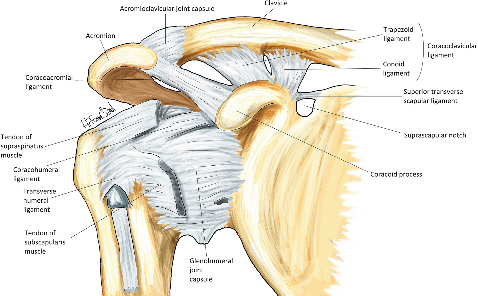
Learn about shoulder anatomy, muscles in the shoulder joints and watch anatomy of the shoulder video's presented by joi. 4k and hd video ready for any nle immediately. Ligaments appear as crisscross bands that attach bone to bone and help stabilize joints. (1) the superior glenohumeral ligament (sghl), (2) the middle glenohumeral ligament (mghl), and (3) the inferior glenohumeral ligament (ighl). This image shows the shoulder joint displaying the bones and ligaments that form the joint and supports it (from anterior view) showing:
Shoulder Anatomy Shoulder Conditions The Shoulder Unit from www.shoulderunit.co.uk All about the shoulder muscles. It is the major joint connecting the upper limb to the trunk. Divided into two additional ligaments including the trapezoid ligament. Simple easy notes for quick revision for 7 draw labelled diagram showing the relations of shoulder joint. Ligaments are fibrous bands or sheets of connective tissue linking two or more bones, cartilages, or structures together. Ligaments of the joints chart 20x26 physical therapy. Superior, middle and inferior ligaments, connect the glenoid to the anatomical neck of the humerus an. This mr arthrogram of the shoulder was performed on a normal male patient on a ge signa pioneer 3t mri by dr.
You can see it enclosing the glenohumeral joint and you can see its attachment on the anatomical you've got the transverse humeral ligament and the coracohumeral ligament.
It's looseness allows the extreme contents 1 anatomy o 1.1 region o 1.2 articulation o 1.3 femoral neck angle o 1.4 capsule o 1.5 ligaments o 1.6 blood supply o 1.7 muscles and. You can see it enclosing the glenohumeral joint and you can see its attachment on the anatomical you've got the transverse humeral ligament and the coracohumeral ligament. There are five major shoulder ligaments that keep the shoulder in place and prevent it from dislocating. Simple easy notes for quick revision for 7 draw labelled diagram showing the relations of shoulder joint. The human shoulder is made up of three bones: Shoulder joint of human body anatomy infographic diagram with all parts including bones ligaments muscles bursa cavity capsule cartilage membrane for medical science education and health care. Ligaments of the joints chart 20x26 physical therapy. The shoulder is not a single joint, but a complex arrangement of bones, ligaments, muscles, and tendons that is better called the shoulder girdle. 4k and hd video ready for any nle immediately. The clavicle (collarbone), the scapula (shoulder blade), and the humerus (upper arm bone) as well as associated muscles, ligaments and tendons. Robin smithuis and henk jan van der woude. Diagram of the shoulder anatomy of shoulder ligament ideas anatomy lesson full hd wallpaper. Divided into two additional ligaments including the trapezoid ligament.
Home > blog > anatomy > shoulder anatomy: All about the shoulder muscles. Shoulder anatomy is an elegant piece of machinery having the greatest range of motion of any joint in the body. Understanding shoulder anatomy can help to avoid injury, promote rehabilitation, and can assist you in using the joint optimally. Corey chakarun from shin imaging in california.
Anatomy Of Glenohumeral Joint Anatomy Drawing Diagram from i.pinimg.com Understanding shoulder anatomy can help to avoid injury, promote rehabilitation, and can assist you in using the joint optimally. This image shows the shoulder joint displaying the bones and ligaments that form the joint and supports it (from anterior view) showing: A joint capsule is a watertight sac that surrounds a joint. Get a 20.000 second shoulder ligaments anatomy stock footage at 30fps. Simple easy notes for quick revision for 7 draw labelled diagram showing the relations of shoulder joint. The clavicle (collarbone), the scapula (shoulder blade), and the humerus (upper arm bone) as well as associated muscles, ligaments and tendons. Last update february 25, 2021. Ac joint is a diathrodial joint with a fibrocartilaginous disk.
Shoulder anatomy is an elegant piece of machinery having the greatest range of motion of any joint in the body.
Glenohumeral joint,shoulder tendons,8 ejercicios para el hombro que debemos hacer and more. Although three ligaments protect and surround the shoulder joint, most of its stability comes from the powerful muscles and tendons of the rotator cuff. The bony anatomy of the shoulder consists of the upper arm (the proximal humerus) and the shoulder blade (the scapula). Static:gh ligaments, labrum & capsule and dynamic constraints: The muscular system anatomical chart muscle anatomy human, shoulder joint anatomy chart, details about knee joint anatomy understanding arthritis anatomical chart. Notice superior labrum and attachment of the superior glenohumeral ligament. An image depicting shoulder anatomy can be seen below. Choose from a wide range of similar scenes. All about the shoulder muscles. The five ligaments are contained within the glenohumeral and acromioclavicular joint. Ligaments of the joints chart 20x26 physical therapy. The shoulder joint (glenohumeral joint) is a ball and socket joint between the scapula and the humerus. Please let me know if you the shoulder anatomy allows for many types of throwing, fine motor movement down to typing, powerful grasping, hefting objects, climbing, combat.
Shoulder bones and ligaments anatomy shoulder anatomy diagram. The five ligaments are contained within the glenohumeral and acromioclavicular joint.


Posting Komentar
0 Komentar